
Introduction
The progressive withdrawal of ventilatory support remains one of the most complex clinical challenges in patients requiring mechanical ventilation in the intensive care unit (ICU).1 Both premature and delayed extubation are associated with increased morbidity, mortality, and prolonged ICU length of stay.2–4 Successful liberation from mechanical ventilation depends on the interaction between recovery from the underlying disease, respiratory muscle strength, the balance between ventilatory load and muscular capacity, and intact central respiratory control.5
Ultrasound has gained a prominent role in critical care because of its portability, immediate availability, and ability to assess respiratory muscle function in real time.6,7 However, its clinical application has largely focused on the diaphragm, generating robust evidence regarding its behaviour during weaning and its ability to predict extubation success. This predominant focus has left the contribution of other respiratory muscle groups, particularly the expiratory muscles, relatively underexplored, despite their important role in ventilatory mechanics.8
During the weaning process, expiratory muscle activity becomes especially relevant. Their activation can reduce transpulmonary pressure, facilitate secretion clearance through an effective cough, and optimise subsequent inspiration by transiently reducing functional residual capacity.9 The elastic recoil generated after active expiration allows inspiratory muscles to initiate the next breath under a lower mechanical load, potentially reducing respiratory effort when ventilatory demand increases.10
Emerging evidence suggests that dysfunction of expiratory muscles is associated with adverse weaning outcomes. Parthasarathy et al.11 demonstrated that extubation failure is associated with increased expiratory muscle activation, reflecting a compensatory response to an unsustainable respiratory load. Similarly, Doorduin et al.12 reported that weaning failure is accompanied by increased expiratory effort and impaired neuromechanical efficiency of the diaphragm. Consequently, expiratory capacity and cough effectiveness have gained clinical relevance and are commonly assessed using peak cough flow in mechanically ventilated patients.13,14
The introduction of ultrasound has enabled direct assessment of abdominal muscle activation during manoeuvres such as coughing. Schreiber et al. showed that a lower abdominal thickening fraction is associated with a higher risk of reintubation and failure to achieve ventilator liberation.15 However, important limitations remain, including high inter- and intra-observer variability and the inclusion of patients with short durations of mechanical ventilation, which limits the generalisability of current findings.8
In this context, a comprehensive synthesis of the available evidence is needed to clarify how abdominal muscle ultrasound has been applied during the weaning process from mechanical ventilation. This review aims to map the existing literature on abdominal respiratory muscle ultrasound in critically ill adults, describing studied populations, ultrasound protocols, measured parameters, and reported weaning or extubation outcomes. Additionally, it seeks to identify methodological heterogeneity, knowledge gaps, and areas requiring standardisation to inform future research and support the development of clinically meaningful assessment frameworks.
Methods
The protocol of this scoping review was developed a priori to ensure methodological transparency and reproducibility and was prospectively registered in the Open Science Framework (OSF) https://osf.io/xnfa5/overview. The review was conducted in accordance with the methodological framework for scoping reviews proposed by the Joanna Briggs Institute (JBI) and is reported following the PRISMA-ScR guidelines.16
Research question (PCC)
To guide the review process and ensure methodological clarity, the research question was formulated using the Population–Concept–Context (PCC) framework, as recommended for scoping reviews.
Population: Adult critically ill patients (≥18 years) receiving invasive mechanical ventilation in the intensive care unit.
Concept: Ultrasound assessment of abdominal respiratory muscles, including the rectus abdominis, internal oblique, external oblique, and transversus abdominis, during the weaning or extubation process.
Context: Weaning from invasive mechanical ventilation in the ICU, including spontaneous breathing trials and pre-extubation assessments, with outcomes related to weaning or extubation success or failure.
Eligibility criteria
This scoping review included studies evaluating adult critically ill patients (≥18 years) receiving invasive mechanical ventilation in the intensive care unit. Eligible studies comprised original research designs (including randomised or quasi-experimental studies, prospective or retrospective cohort studies, case–control studies, and analytical cross-sectional studies with follow-up) that applied ultrasound assessment of abdominal respiratory muscles (rectus abdominis, internal oblique, external oblique, and/or transversus abdominis) during the weaning or extubation readiness process, including spontaneous breathing trials or pre-extubation assessments.
Studies were required to report ultrasound-derived measurements of abdominal muscle structure or function, such as muscle thickness or thickening fraction, in relation to weaning or extubation processes or outcomes. Full-text availability was required. No restrictions were applied regarding publication date, language, or geographical region.
Studies were excluded if they involved paediatric populations, were conducted outside the ICU, focused exclusively on diaphragmatic or lung ultrasound without abdominal muscle assessment, or were non-original research (case reports, small case series, narrative or systematic reviews, editorials, study protocols, conference abstracts lacking sufficient data), as well as animal or simulation studies.
Search strategy
Two investigators (HMPG, LAPL) independently conducted a comprehensive literature search in PubMed, Embase (Ovid), the Cochrane Library, Scopus, Web of Science, ScienceDirect, and Epistemonikos, from database inception to January 2026. Following peer-review, an updated search was performed on May 30, 2026, to identify potentially eligible studies published after the original search date. This updated search identified one additional eligible study, which was subsequently included in the review. Discrepancies were resolved by consensus and, when necessary, through consultation with a third reviewer.
In addition, manual searches of the reference lists of included articles, forward and backward citation tracking, and searches of grey literature sources, including thesis and dissertation repositories, were performed to identify potentially relevant studies not captured through electronic database searches.
The search strategy combined Medical Subject Headings (MeSH) terms and free-text keywords related to ventilator weaning and abdominal muscle ultrasound, including “mechanical ventilation”, “weaning”, “extubation”, “ultrasound”, “abdominal muscles”, “rectus abdominis”, “oblique muscles”, and “transversus abdominis”, as well as free-text terms such as “abdominal muscle thickening fraction” and “abdominal ultrasound for weaning”. No language restrictions were applied. The complete search strategy is provided in Supplementary File 1.
Data synthesis and extraction
A structured data extraction form was used to systematically and consistently collect information across included studies. For each article, the following variables were extracted: first author, year of publication, country, study design, sample size, causes of mechanical ventilation, duration of mechanical ventilation prior to assessment, abdominal muscles evaluated (rectus abdominis, internal oblique, external oblique, transversus abdominis), ultrasound parameters measured (e.g., muscle thickness or thickening fraction), timing of the assessment (tidal breathing, coughing, or spontaneous breathing trial), and definitions of weaning or extubation outcomes as reported by the authors. When available, proposed cut-off values related to weaning or extubation outcomes were also recorded.
Data extraction was performed independently by two reviewers (HMPG, LAPL) using Rayyan,17 A pilot test was conducted on a subset of studies to refine the extraction form and ensure consistency. Extracted data were summarised descriptively and organized to support a narrative synthesis aimed at mapping study characteristics, ultrasound methodologies, and reported outcomes.
Risk of bias assessment
In line with the exploratory objectives of this scoping review, an assessment of methodological limitations was undertaken to describe the quality and heterogeneity of the available evidence, rather than to inform study exclusion or quantitative synthesis. The methodological characteristics of the included studies were evaluated using the QUADAS-2 tool,18 which assesses the domains of patient selection, index test, reference standard, and flow and timing.
Although formal risk-of-bias assessment is not a mandatory component of scoping reviews, the included studies evaluated ultrasound-derived parameters as index tests in relation to clinically relevant outcomes (weaning or extubation success or failure). Therefore, QUADAS-2 was considered an appropriate framework to systematically characterise methodological limitations, potential sources of bias, and applicability concerns across studies. The assessment was conducted independently by two reviewers. Its findings were used solely to contextualise the mapped evidence and identify methodological gaps, and were not used to determine study eligibility, exclude studies, or grade the certainty of the evidence.
Results
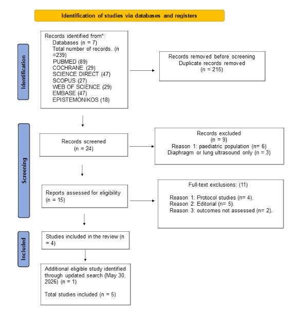
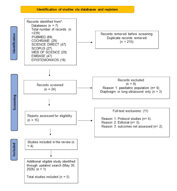
The initial search identified 239 records across seven databases (PubMed, the Cochrane Library, Scopus, Web of Science, ScienceDirect, Embase, and Epistemonikos). After removal of 215 duplicate records, 24 unique records remained for title and abstract screening. Of these, nine records were excluded because they involved paediatric populations or did not include abdominal muscle ultrasound assessment. A total of 15 full-text articles were assessed for eligibility, of which 11 were excluded because they were protocol studies, editorials, or did not address the concept of interest. Following an updated search conducted on May 30, 2026, one additional eligible study was identified and included. Ultimately, five studies met the inclusion criteria and were included in this scoping review. A detailed flow diagram of the study selection process is presented in Figure 1.
Characteristics of the studies included
The characteristics of the included studies are summarised in Table 1. The studies were published between 2021 and 2026. All five included studies employed prospective observational designs.15,19–22 Across the included studies, the indications for initiating mechanical ventilation varied considerably. Three studies15,19,21 evaluated mixed ICU populations in whom mechanical ventilation was required for heterogeneous medical, surgical, and infectious causes, including respiratory failure due to sepsis, pneumonia, cardiac failure, postoperative complications, and other critical conditions of diverse aetiology.
In contrast, one study20 focused exclusively on patients with neurological critical illness, including stroke, traumatic brain injury, and postoperative neurosurgical conditions. In this population, neurological dysfunction was the primary driver of dependence on mechanical ventilatory support, which may influence patterns of abdominal respiratory muscle activation during the weaning process. Another study22 evaluated patients with spinal trauma, including individuals with and without spinal cord injury, in whom respiratory muscle dysfunction and altered respiratory mechanics may influence ventilator liberation outcomes.
The definition of weaning or extubation success varied across the included studies. Qiu et al.20 defined extubation success as the absence of reintubation within 48 hours following tube removal. Schreiber et al.15 applied a broader criterion, considering success as the absence of reintubation or reconnection to mechanical ventilation within 72 hours after extubation. More recently, Schreiber et al.22 defined weaning failure at the first attempt as reintubation after extubation, inability to achieve extubation, or death while still receiving mechanical ventilation. In contrast, the studies by Amara et al.19 and Bansal et al.21 did not provide an explicit definition of weaning or extubation success, which limits direct comparability across studies. Nevertheless, all investigations assessed outcomes related to the sustained tolerance of spontaneous breathing after withdrawal of invasive ventilatory support.
Information on operator background and ultrasound expertise was reported inconsistently across studies (Table 2). Where available, ultrasound assessments were performed by trained healthcare professionals, including a respirologist, a respiratory therapist, and a physiotherapist with specialized ultrasound training and clinical experience. However, the remaining studies provided limited or no information on operator qualifications, ultrasound expertise, or whether assessments were performed by routine clinical staff or dedicated research personnel.
Duration of mechanical ventilation prior to assessment
Two studies reported a ventilation duration of 48 hours at enrolment,20,21 one did not report this variable ,19 and one described a longer duration of 4–5 days before ultrasound assessment ,15 and one performed serial assessments during mechanical ventilation, with a median of 12 days from intubation to the final measurement before the first weaning attempt.22
Ultrasound assessment protocols
All studies applied ultrasound to assess abdominal respiratory muscles, but substantial methodological heterogeneity was observed. Assessments were performed during tidal breathing, voluntary coughing, or at specific stages of the SBT. One study conducted serial measurements throughout the SBT,15 whereas the others relied on single-point evaluations. In contrast, Schreiber et al.22 performed longitudinal ultrasound assessments two to three times weekly from ICU admission until the first weaning attempt, ICU discharge, or death. Differences were also noted in patient positioning, respiratory phase, and measurement sequence, underscoring the absence of a standardized ultrasound protocol during weaning.
Ultrasound parameters
All studies assessed abdominal muscle thickness (rectus abdominis, internal oblique, external oblique, and transversus abdominis) as a structural parameter. In contrast, dynamic functional assessment was less consistent; three studies measured abdominal muscle thickening fraction, including assessments during voluntary coughing and longitudinal monitoring of respiratory muscle activity.15,20,22 Two studies additionally incorporated diaphragmatic ultrasound indices, including excursion and diaphragmatic thickening fraction.19,21 No unified analytical framework or externally validated cut-off values were applied across studies.
Comparison between successful and failed weaning/extubation
Across studies, patients who failed weaning or extubation generally showed reduced abdominal muscle performance. Amara et al.19 reported lower abdominal muscle thickness in patients with difficult or prolonged weaning, with moderate discriminative performance (AUC 0.689–0.718). Proposed cut-offs included 0.638 cm for the rectus abdominis, 0.492 cm for the internal oblique, 0.315 cm for the external oblique, and 0.253 cm for the transversus abdominis. Diaphragmatic excursion showed moderate discrimination (AUC 0.700), whereas diaphragmatic thickening fraction had limited value (AUC 0.548).
In neurocritical patients, Qiu et al.20 reported marked differences in dynamic abdominal muscle recruitment. Internal oblique thickening fraction during voluntary coughing demonstrated the highest discriminative performance (AUC 0.957), with a threshold ≥ 34.15% yielding a sensitivity of 93.8% and specificity of 75%. Similar findings were observed for the external oblique.
Bansal et al.21 reported more modest discrimination, with an AUC of 0.64 for the internal oblique and limited performance for the external oblique (AUC 0.55) and transversus abdominis (AUC 0.44). In the bi-centre study by Schreiber et al.15, reduced abdominal thickening during voluntary coughing was associated with ventilator liberation failure; each 10% decrease in thickening fraction increased the odds of reintubation (OR 2.1; 95% CI 1.1–4.4). Similarly, Schreiber et al.22 reported that patients who failed weaning exhibited a greater reduction in internal oblique thickness over time, whereas successful patients maintained higher muscle thickness values during the observation period.
Overall, dynamic abdominal muscle assessment, particularly thickening fraction during coughing, showed greater discriminative capacity than static thickness alone. Patients with weaning or extubation failure exhibited both reduced muscle thickness and impaired contractile activity.
Assessment of the methodological quality of the studies
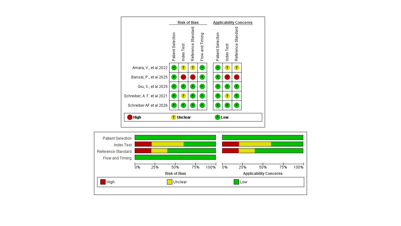
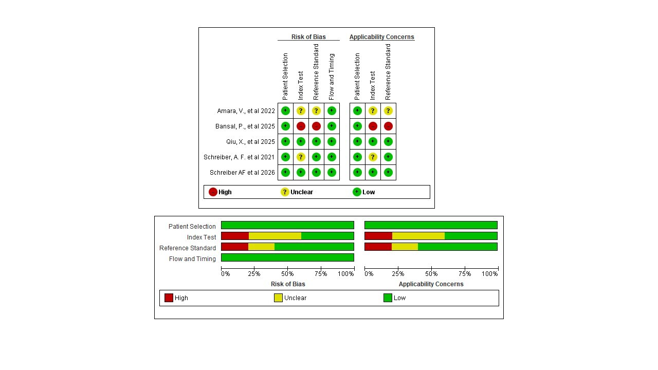
QUADAS-2 revealed heterogeneous methodological quality, mainly within the reference standard domain. Three studies were judged to be at low risk of bias owing to clearly defined weaning or extubation outcomes and consistent assessment procedures.15,20,22 In contrast, Bansal et al.21 was rated at high risk of bias because no explicit definition of weaning or extubation success was provided, affecting both the reference standard and applicability domains. Amara et al.19 demonstrated a moderate risk of bias for similar reasons.
Across all studies, the index test and flow and timing domains were consistently rated as low risk of bias. Regarding applicability, concerns were highest for Bansal et al.21 and moderate for Amara et al.,19 whereas no major applicability concerns were identified in the remaining studies. A visual summary of the QUADAS-2 assessment is presented in Figure 2.
Discussion
This scoping review identified five prospective observational studies comprising a total of 233 adult ICU patients receiving invasive mechanical ventilation. The mapped evidence indicates that selected ultrasound-derived parameters of the abdominal respiratory musculature, particularly muscle thickness and cough-related thickening fraction, have been explored as markers of a patient’s ability to tolerate spontaneous breathing following withdrawal of invasive ventilatory support, including through longitudinal assessment of respiratory muscle changes over time. However, the interpretation of these findings is constrained by substantial methodological heterogeneity across studies.
Marked variability in ultrasound acquisition protocols, inconsistency in definitions of weaning or extubation success, differences in the timing and frequency of ultrasound assessments, and limited sample sizes precluded quantitative synthesis and hindered the development of robust or generalizable diagnostic frameworks. Rather than supporting definitive clinical decision-making, the available evidence primarily reveals exploratory signals and highlights important methodological gaps that must be addressed before abdominal muscle ultrasound can be reliably integrated into routine weaning assessment.
To our knowledge, this is the first scoping review to systematically map the existing literature on the use of abdominal muscle ultrasound during weaning or extubation from mechanical ventilation in critically ill adults. By characterizing study designs, ultrasound methodologies, assessed parameters, and reported outcomes, this review provides a structured overview of the current state of the field and identifies key areas requiring standardization and further investigation.
Across the included studies, a recurring pattern was observed in which patients who experienced weaning or extubation failure tended to exhibit reduced abdominal muscle thickness and lower thickening fraction values. This observation is physiologically consistent with the established role of the expiratory muscles in generating abdominal pressure to support an effective cough. Notably, dynamic ultrasound-derived parameters, particularly cough-related thickening fraction, were more frequently associated with weaning outcomes than static thickness measurements alone, although longitudinal reductions in abdominal muscle thickness were also associated with weaning failure in some studies, suggesting that abdominal contractile activity has been preferentially explored as a marker of respiratory muscle reserve during the weaning process.
Nevertheless, these findings must be interpreted within the context of weaning as an inherently multifactorial process. Respiratory musculature represents only one component of a complex physiological system, and successful liberation from mechanical ventilation depends on the interaction of multiple factors, including pulmonary parenchymal function and overall respiratory mechanics, neurological status and coordination of cough, airway protection, and haemodynamic and metabolic stability.23 Accordingly, abdominal muscle ultrasound should be viewed as a complementary component within a broader, multimodal assessment strategy rather than as an isolated determinant of weaning success.
Within this framework, cough is widely recognized as a key determinant of extubation outcomes, particularly regarding airway protection and patency. Several studies have demonstrated that reductions in cough peak flow and tussive strength are associated with an increased risk of reintubation.10,14,24 As the generation of adequate cough peak flow largely depends on effective recruitment of the expiratory musculature, ultrasound assessment of the abdominal muscles has been explored as a means of providing complementary information on cough-related function and airway-protective capacity following endotracheal tube removal. Importantly, such assessment should not be considered in isolation but rather interpreted within an integrated clinical framework that accounts for respiratory mechanics, neurological status, haemodynamic stability, and the coordinated interaction between the diaphragm, expiratory muscles, and the broader respiratory system.
Traditionally, objective evaluation of cough function in critically ill patients has relied on measures such as maximal expiratory pressure (MEP) and cough peak flow (CPF).25,26 Although these tools are well established, they depend heavily on voluntary patient cooperation, limiting their applicability in individuals with impaired consciousness, neuromuscular dysfunction, or reduced ability to follow commands. In such circumstances, an important gap persists in the objective assessment of tussive function.
Within this context, ultrasound assessment of the abdominal musculature has been proposed as a potential alternative approach, as it does not require maximal voluntary effort and allows evaluation of both muscle structure and activation during provoked or assisted coughing manoeuvres. Measurement of abdominal muscle thickening fraction during coughing may therefore provide indirect insight into expiratory muscle engagement and, by extension, cough-related function, particularly in patients for whom conventional volitional testing is not feasible. From the perspective of this scoping review, abdominal ultrasound may thus be considered a complementary component of cough assessment in selected critically ill patients, while acknowledging the exploratory nature of the current evidence.
When synthesizing findings across the five included studies, the internal oblique (IO) muscle emerged as the most consistently reported abdominal parameter. Both static thickness measurements19,21 and dynamic assessments of cough-related thickening fraction15,20 more frequently demonstrated differences between patients who successfully weaned or were extubated and those who did not. Among the reported parameters, the IO muscle was associated with the highest discriminative values, with AUC estimates reaching up to 0.957 in selected analyses.
Despite these associations, the available studies do not permit the establishment of a causal relationship between abdominal muscle dysfunction and weaning failure. Reductions in abdominal muscle thickness or thickening fraction may reflect broader processes such as global muscle weakness, critical illness–associated neuromuscular dysfunction, or greater overall disease severity, rather than an isolated mechanism directly responsible for failure of ventilator liberation. Accordingly, these findings should be interpreted as associations rather than evidence of direct causality.
Methodological heterogeneity across ultrasound protocols represents a major limitation for evidence synthesis. Variability in measurement timing, frequency of assessment, the manoeuvre performed (rest, spontaneous breathing, or coughing), patient positioning, and the selection of ultrasound-derived parameters (thickness versus thickening fraction) are all factors capable of influencing abdominal muscle behaviour and measurement results. This heterogeneity likely contributes to discrepancies between studies and underscores the need for standardized acquisition and analysis protocols for abdominal muscle ultrasound during ventilatory weaning. Although dynamic parameters, particularly cough-related thickening fraction, appear physiologically relevant, their clinical applicability cannot be consolidated without greater technical and operational uniformity.
The limited number of studies identified in this review may partly reflect these methodological challenges. Unlike diaphragmatic ultrasound, which benefits from relatively standardized acquisition protocols and widely accepted parameters such as diaphragmatic excursion and thickening fraction, the assessment of abdominal respiratory muscles remains heterogeneous. Different studies have evaluated distinct muscle groups, anatomical landmarks, respiratory manoeuvres, and ultrasound-derived variables, while universally accepted reference values and standardized assessment protocols are still lacking. These factors may have hindered reproducibility across studies and limited the broader adoption of abdominal muscle ultrasound in both clinical practice and research.
In addition, ultrasound measurement protocols varied substantially with respect to anatomical measurement sites and probe orientation. Limited reporting of patient positioning and transducer stabilization during manoeuvres such as coughing further restricts reproducibility. Small anatomical variations may significantly affect thickness and thickening fraction values,27,28 reinforcing the importance of standardization in future investigations.
Although none of the included studies directly correlated abdominal muscle thickening fraction with cough-related measures such as cough peak flow or maximal expiratory pressure in critically ill patients, evidence from other populations suggests potential associations between ultrasound-derived muscle activity and expiratory capacity. In healthy individuals, Schreiber et al. demonstrated correlations between abdominal muscle thickening fraction and pressure generation during expiratory efforts and coughing.15 Similarly, Ishida et al.28 reported moderate correlations between abdominal muscle thickness, particularly of the internal and external oblique muscles, and peak expiratory flow in young adults and older women. However, these associations, derived largely from non–critically ill populations, are insufficient to support substitution of established measures such as MEP or CPF, and instead support the role of abdominal ultrasound as a complementary assessment tool in selected clinical contexts.
Notably, the highest discriminative values for abdominal muscle thickening fraction, particularly involving the internal oblique muscle, were reported in neurocritical patients. In this population, where impairment of neuromuscular control and cough effectiveness is common29
, ultrasound-based evaluation of expiratory musculature may offer greater relative value than in general ICU populations. This observation highlights the importance of future studies stratified by patient phenotype, neurological status, and underlying mechanisms of ventilator dependence.
Overall, the methodological assessment identified variability across studies, with the main concerns related to the reference standard domain and incomplete reporting of ultrasound procedures. These findings highlight important methodological limitations and reinforce the need for greater standardization in future studies evaluating abdominal muscle ultrasound during ventilator weaning, including the timing, frequency, and technical execution of ultrasound assessments.
From a clinical perspective, abdominal muscle ultrasound may be considered a complementary component of weaning assessment in selected scenarios, particularly in patients at increased risk of ineffective cough or with limited ability to cooperate with volitional testing. Its potential value lies in integration within a multimodal evaluation strategy rather than as a standalone substitute for established clinical criteria.
Strengths and limitations
A key strength of this scoping review is its focused examination of abdominal muscle ultrasound during weaning from mechanical ventilation, an area that remains underexplored in critical care practice. The review followed established methodological standards for scoping reviews, including a comprehensive search across multiple databases and a structured approach to data extraction and descriptive synthesis. Use of a validated tool to characterize methodological limitations further enhanced transparency in mapping study quality and heterogeneity.
Several limitations warrant consideration. The number of available studies was small, sample sizes were limited, and substantial heterogeneity was observed in ultrasound acquisition protocols, timing of measurements, frequency of assessment, and definitions of weaning or extubation success. These factors precluded quantitative synthesis and limit the robustness and generalisability of cross-study comparisons. Moreover, the observational nature of the included studies does not permit causal inference, and the absence of external validation for proposed ultrasound cut-off values restricts immediate clinical applicability. Finally, although grey literature was searched, publication bias cannot be excluded, as the limited number of studies prevented a formal assessment of small-study effects.
Implications for future research
Future studies should prioritize the development and validation of standardized abdominal muscle ultrasound protocols, including clear definitions of measurement timing, manoeuvres, and anatomical landmarks, and longitudinal assessment strategies. In addition, investigations should assess the incremental value of abdominal ultrasound relative to established weaning assessment tools and explore its potential impact on clinically relevant outcomes such as extubation failure, duration of mechanical ventilation, and ICU length of stay. Particular attention should be given to specific patient subgroups, such as neurocritical populations, in whom assessment of cough and expiratory muscle function is especially challenging.
Conclusion
Ultrasound assessment of the abdominal respiratory muscles has been explored as a source of complementary information during the weaning process from mechanical ventilation. However, the currently available evidence is limited by small sample sizes, substantial heterogeneity in assessment protocols, and inconsistent definitions of weaning or extubation success. As a result, no single ultrasound-derived parameter can be considered sufficiently consistent to support routine clinical application at this stage. Future research should focus on developing standardized acquisition protocols and conducting well-designed studies to clarify the potential role of abdominal muscle ultrasound in predicting weaning and extubation outcomes.
Acknowledgements
The authors would like to acknowledge Claudia Ruiz, Head of the Department of Therapy and Rehabilitation at Clínica Reina Sofía, for her valuable support and encouragement of this research.
Availability of data and materials
The datasets used and/or analyzed during the current study are available from the corresponding author on reasonable request.
Competing interests
The authors declare that they have no competing interests.
Funding
This study did not receive any specific grant from funding agencies in the public, commercial, or not-for-profit sectors.
Authors’ contributions
HMPG, LAP, LAPL, DME, and DMF contributed to the study conceptualization, methodology, data analysis and interpretation, as well as to the drafting and revision of the manuscript. HMPG and LAP critically reviewed the statistical methods. All authors contributed substantially to manuscript editing and revision, read the final version, and approved it for submission.
Ethics approval
Not required for this article type.
AI Statement
The authors confirm that no generative AI or AI-assisted technology was used to generate the scientific content of this manuscript. AI-assisted tools were used only for language editing and improvement of clarity.