Introduction
Point-of-care ultrasound (PoCUS) refers to a portable ultrasound investigation performed and interpreted at the bedside by a healthcare provider. The first of its two main uses is to assist with procedure guidance.1 Ultrasound (US)-guided central venous catheter placement is now considered the standard of care2; however, PoCUS can also be used to guide low-risk procedures, such as peripheral intravenous (PIV) cannulation. PoCUS-guided PIV catheter insertion is associated with an improved success rate, reduced number of punctures, faster procedure, and, most importantly, increased patient satisfaction, all without any increased adverse events.3,4 The same themes hold true when PoCUS is used to guide the insertion of a radial arterial catheter.5 The second role for PoCUS is to answer focused clinical questions through supplementing the physical exam. In fact, the recent Society of Critical Care Medicine guideline on use of critical care ultrasound recommends using PoCUS to aid with the diagnosis and management of adults admitted to hospital with acute respiratory failure.6 Compared to conventional approaches, PoCUS reduced: 1) the time to correct diagnoses; 2) the time to appropriate treatment administration; and 3) the duration of mechanical ventilation.6
While early PoCUS adopters were predominantly physicians,7,8 many other health professionals (such as nurses9 and physiotherapists10) have started to use it in their practice. As of the 2010s, respiratory therapists (RTs) have begun incorporating this tool into practice,11 and PoCUS training for this profession is gaining momentum. A recent scoping review performed by our group identified nine published PoCUS training protocols for RTs in North America, Europe, South America, and Asia. The majority of educational techniques used a combination of didactic talks, hands-on training, and practical assessment.11 Of note, the courses usually offered the training to a small group of participants, ranging from 2 to 100.
As such, we undertook the task of creating a course to deliver PoCUS education to Canadian RTs on a national scale. Herein, we describe the course’s development, implementation, and evaluation, as well as participants’ perceptions of the course and their self-reported PoCUS confidence and knowledge.
Methods
Course delivery
Course set-up
The course took place annually at the Canadian Society of Respiratory Therapists (CSRT) Conference from 2023-2025 (2023: Charlottetown, Prince Edward Island; 2024: Banff, Alberta; 2025: Ottawa, Ontario). A total of three courses were offered through the CSRT. Two smaller, local courses were also offered out of St. Joseph’s Healthcare Hamilton for local RTs who were unable to attend the conference, one in 2024 and one in 2025. Pre-registration was required to attend, as capacity was limited to ensure there was ample time for hands-on practice. All participants were provided with didactic material to review prior to attending.
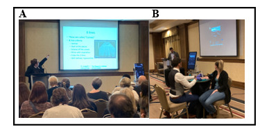
The logistics of the one-day sessions were as follows: A large room was arranged with chairs for participants in the middle of the room, with a podium, microphone, projector, and laptop at the front to deliver the introductory didactic lecture (Figure 1A and 1B). Six stations were placed around the periphery of the room to facilitate a smooth transition to the hands-on small group session. Each of the six stations was labelled as #1-6, equipped with an US machine, US gel, hand sanitizer, and paper towel for clean-up. Four of the six stations had standardized patients, and the remaining two stations had US task trainers, an assortment of vascular access catheters, and sharps disposal containers.

Course instructors
The course instructors were intentionally multidisciplinary. Each year, the course was led by an adult critical care physician (KL), an adult RT clinical resource leader (KH), an adult anesthesia assistant (AA) (JB), a pediatric AA (BD), and a pediatric anesthesiologist (MK). Other physicians ran one of the small groups (e.g., adult critical care physicians [KLC and JR] or a diagnostic radiologist [EZ]). The team members were from either McMaster or Dalhousie University.
Course standardized patients
The standardized patients (SPs) were crucial for the success of the small group hands-on stations, where participants practiced landmarking and scanning on real people. Standardized patients were typically RT students, local RTs/AAs, or RTs/AAs who were conference participants. Two of the RTs were supported by their respective industry employers to participate as standardized patients.
Ultrasound machines
Ultrasound machines were provided on loan by several companies that were exhibiting at the conference. There was a wide variety of different US brands present (i.e., Mindray, GE, and Butterfly) to ensure generalizability and increased comfort across many brands, acknowledging that every RT may have access to a different US machine at their home institution.
Introductory lecture
An hour-and-a-half lecture was delivered at the start of the course. The objective of the lecture was to provide the basic information required to perform a lung PoCUS and PoCUS-guided vascular access. After the moderator’s welcome and instructor introductions, there was a review of the key points, such as the definition and purpose of PoCUS, transducer selection, “knobology”, and exam pre-sets. For the lung PoCUS portion of the talk, landmarking, patient positioning, image acquisition, and lung signatures, including A lines, B lines, pleural effusions, consolidations, and lung sliding, were all taught. This was followed by the vascular access portion of the lecture, which addressed the benefits and indications for PoCUS-guided access, reviewed upper-extremity venous and arterial anatomy, and discussed image acquisition techniques and dynamic needle-tip positioning. A live demonstration of a PoCUS-guided PIV catheter insertion was also done. The session concluded with a review of the supporting evidence for the efficacy and safety of PoCUS-guided vascular access.
Breakout hands-on small group sessions
Participants were divided equally (five to eight participants per station) among the six stations. Upon entering the course, each participant received an envelope containing a number corresponding to one of the stations. Each participant began the hands-on small-group session at the assigned station.
Stations one through four were related to lung PoCUS, and stations five and six were related to US-guided intravascular access (Table 1). Participants spent 25 minutes at each station working on the specified learning objectives. The moderator of the session kept time and announced when there were 5 minutes remaining at a station and when it was time for participants to rotate to the next station in a clockwise manner.
Large group wrap-up
With the remaining five minutes, all participants moved back to the middle of the room for a wrap-up large group lecture. Here, a summary was provided, tips and tricks on how RTs could implement PoCUS education at their local hospitals, and resources were shared. Participants then completed the post-session survey.
Survey
Development of the survey
A survey (please see Supplementary Figure 1) was developed by the core group of instructors (KL, KH, BD, and MK). In total, there were 31 questions. The item generation was done through a literature review on the topic,11 and discussion for item reduction. The following themes were identified as pertinent and ultimately included on the survey:
-
Six questions on participants’ relevant demographics (including province of current practice, years in practice, type of primary clinical practice, designations, and if the participant has access to an US machine at their home institution).
-
Four questions on the participant’s current use of PoCUS for vascular access, diagnostics, and any prior formal training in PoCUS.
-
Twelve total questions on the participant’s perceived knowledge of PoCUS (transducer selection, image acquisition, image interpretation, and vascular cannulation) before and after the course.
-
Nine items to provide feedback on the course delivery (level of difficulty, duration, pacing, methods of instruction, etc.).
All survey questions utilized a five-point Likert scale from strongly disagree to strongly agree (or similar, depending on the question) to allow for generalized responses.
Survey distribution
The surveys were distributed on paper found within the participant’s envelope, which they had received at the beginning of the course. Participants were given time at the end of the wrap-up session to complete the survey. Participants were asked to return the survey to one of the instructors or moderator as they left the room to encourage maximal participation.
Of note, as this course (and hence survey) was delivered multiple times over a three-year period, there were slight variations in the survey questions, so not all questions have the same number of responses, as some questions were not included in earlier iterations. For example, the vascular access lecture and stations were only offered in the second and third year of course delivery.
Data analysis
Survey data were exported into Microsoft Excel (version 16.78, Redmond, WA, USA), and all statistical analyses were conducted using Microsoft Excel. The data are presented categorically, by domain and in aggregate. Descriptive statistics are calculated as frequencies and percentages, as all data is categorical. Given the anticipated small number of responses, we elected to only use descriptive statistics.
Ethics
Given that the collection of this information would be regularly collected and shared by CSRT as part of their conference evaluations, research ethics board approval was not needed.12 This survey would fall under the umbrella of quality assurance. The survey did not collect any identifiable information. Participants were able to voluntarily withdraw at any point in time with no repercussions.
Results
Participant demographics
Participants enrolled in the course via the conference registration system, with spots allocated on a first-come, first-served basis. The course was delivered to a total of 117 participants over five courses (Table 2). The majority of participants were from Ontario (55%), followed by Alberta and British Columbia. Most participants were in their first five years of practice or in years 11-20. Allowing for multiple answers, over 50% of participants had their primary practice in a university-affiliated teaching hospital. Seventy-seven percent of participants were registered respiratory therapists (RRTs) and 14% were student RTs. Seventy-one respondents (71%) practiced in critical care (adult, pediatric, or neonatal), 23 (18%) practiced in the operating room, and the remaining 11% practiced in primary care, rehabilitation, or other locations.
Prior experience with PoCUS
The vast majority (90%) of respondents had access to a US machine at work, 5% did not, and the remaining 5% were uncertain (see Supplementary eTable 1). Approximately 90% of respondents had not previously attended a formal course on either PoCUS-guided vascular access or lung PoCUS. In keeping with the lack of training, 73% of respondents had never performed a bedside lung PoCUS, and 50% had never (or only once) used PoCUS to guide vascular access.
Knowledge before and after the course
Transducer selection
Participants reported that their overall knowledge improved after taking the course (Table 3). For instance, 27% of participants agreed or strongly agreed that they could select the correct transducer prior to the workshop, compared to 89% after. Likewise, 50% agreed or strongly agreed they could select the correct transducer to guide vascular access before the course, compared to 95% after.
Image acquisition
Participant comfort in image acquisition also improved. Those who agreed or strongly agreed they could obtain a good image for lung PoCUS before the course were 17% compared to 84% after. Vascular access image acquisition comfort rose from 54% agreeing/strongly agreeing they could acquire a good image to 93% after the course.
Image interpretation
Likewise, participants were far more likely to agree/strongly agree that they could interpret either a lung PoCUS (70% vs 10%) or use PoCUS to guide vascular access (87% vs 48%) after the course, compared to before.
Course feedback
Lecture
Every participant (n=117, 100%) who took the course felt that the lecture was at an appropriate level of difficulty (Table 4). The vast majority (87%, n=102 participants) felt that the review lecture was a perfect speed, while 12% (n=14) found it a bit too fast.
Hands-on small group sessions
Many participants felt that the time for the small group hands-on sessions was perfect (80%, n=94 people), while 16% found it a little too short and 3% found it too long. Overall, the vast majority found the standardized patients (96%) and the vascular access models (97%) to be very helpful in consolidating learning.
Overall feedback
Most people found the workshop to be an appropriate duration (81%), with the remaining participants finding it either too short or too long in an even distribution. Ninety-nine percent either agreed or strongly agreed that receiving the workshop from an interdisciplinary team was enjoyable. Lastly, all participants (100%, n=117) would recommend the workshop to a colleague.
Discussion
In this survey of 117 participants in the CSRT PoCUS course, we explored participants’ reported knowledge pre- and post-course, as well as their overall course feedback. We found that most participants gained self-reported comfort and knowledge in transducer selection, image acquisition, and image interpretation for both lung PoCUS and PoCUS-guided vascular access. Most found the level of difficulty appropriate and the hands-on small-group sessions helpful for consolidation. The duration of the course and all components were stated to be appropriately timed, although some participants reported the small-group hands-on session at 25 minutes to be too short.
We believe that we have demonstrated that our program is a feasible, well-received, practical course for RTs to gain preliminary confidence and competence with lung PoCUS and PoCUS-guided vascular access (PIV and radial arterial lines). In fact, this course design is similar to components of the PoCUS curriculum delivered to internal medicine residents at McMaster University.13 However, practice, repetition, and ongoing consolidation is unequivocally necessary for anyone learning PoCUS. In our scoping review, we found that 86% of PoCUS programs for allied healthcare workers required a practical assessment of knowledge, often including a median of 11 reviewed scans.11 The need for repetition was demonstrated in a recently published curriculum where 10 Canadian RTs were taught lung PoCUS.14 RTs were required to undergo a didactic lecture followed by a 3h hands-on training session. Over the next 6 weeks, they were required to perform 10 lung PoCUS scans. Approximately 85% of RTs were able to acquire and correctly interpret their images, although only 2 RTs completed the additional scan requirements. The Canadian recommendations for “critical care ultrasound training and competence” require physicians to perform 20 scans, with the scans reviewed and critiqued.15 As such, it is necessary that, should a hospital opt to permit RT PoCUS studies, there should be a local champion who can continue education after our initial teaching.
We received some comments that the hands-on small-group session was too short. While it is certainly expected that some participants will require more coaching and one-on-one time to acquire appropriate images, we believe there may be another explanation for that finding. In the last course we ran, participants who failed to register for the session may have attended. This likely caused more participants to attend each small group session than we had anticipated, resulting in not all participants having a long enough turn. While ideally everyone who wants to take the course will be permitted to do so, we may have to move away from the honour system and check pre-registration confirmation at the door in the following years, and possibly run more courses during the conference to accommodate demand.
Our study has important strengths. First, the course was delivered to over 100 different RTs over a three-year period, demonstrating feasibility and ongoing interest by RTs. This is a course that would be easily adapted to any local hospital, provided there are PoCUS experts who are willing to champion the RTs. This was demonstrated by the fact that we performed two smaller local courses, delivering the same curriculum. Moreover, our survey has demonstrated a substantial increase in perceived confidence and knowledge of both lung PoCUS and PoCUS-guided vascular access immediately after the course.
There are limitations to our study. First, the survey assessed self-perceived knowledge and confidence. Given that this course was administered over almost an entire day at a conference by unfunded instructors, we did not allot additional time for a written or practical exam to assess actual competence. The personnel resources required to conduct a post-course exam would be infeasible and would require substantial financial support. In addition, it would be quite atypical for this type of course to have a formal exam. As mentioned above, the course did evolve over time. Our first year of delivery in Charlottetown did not include the vascular access section-hence the alterations to the survey and having a varying number of survey responses. In addition, the survey did evolve to ask improved questions, although given the large number of respondents, 100% response rate, and consistent overall trends, our data likely demonstrates reliability. We believe we have a 100% response rate. It is possible that a participant may not have handed in their envelope with the survey upon exit of the course; however, this is unlikely to account for significant variation in numbers or findings. Lastly, we did not undertake comparative statistics due to the survey variation, as well as the anticipated low number of overall responses.
The largest criticism of our study is that this course is only one step forward in a journey to become a competent PoCUS provider. As indicated above, it is important that RTs identify a local champion in PoCUS to help grow their knowledge. For example, at St. Joseph’s Healthcare in Hamilton, some RTs and AAs have become local champions through continued mentorship from critical care physicians and anesthesiologists. In fact, one of the RTs has gained a high skill level and has taught resident physician PoCUS education sessions. Another example is at the IWK Health Centre, where site champions (a core group of pediatric anesthesiologists) have trained the pediatric AA group to be “superusers” with ultrasound for vascular access. The AA team now provides the majority of PICC and challenging access services throughout the facility for neonatal, pediatric, and adult patients. These AAs also teach vascular access PoCUS to nurses, physicians, allied healthcare staff, and learners within this institution and beyond. We acknowledge that it may be easier to find PoCUS champions in academic as opposed to community centres. An interesting next step would be to follow up with future course attendees over the longer term (i.e., 6 months and 1 year post-course) to see whether they have had the opportunity to further their skills in the clinical setting and whether they are confident in their retention of the key information. As of present, this only evaluates our program at the Kirkpatrick level one (assessing engagement, relevance, and satisfaction). Further follow-up done at the home institute would assess Kirkpatrick levels two through four.16
It is reasonable to consider that in the future, formal, high-quality PoCUS training will exist in the curriculum for RTs, AAs, and other allied health professionals. This may, however, take time and require collaboration between stakeholders such as governing/accrediting bodies, educational institutions, researchers, and others. At present, the power of PoCUS continues to be harnessed at the bedside across acute care areas, with increased evidence in favour of its use, availability of machines, and clinician training. This course has modelled an interdisciplinary approach to teaching PoCUS with very satisfactory short-term results. It may be possible to adopt this model and scale it to local needs. This may be offered to both students and practicing clinicians striving to meet current clinical expectations. As discussed, implementation success hinges on site champions where learners work, to facilitate ongoing skill development, foster safe learning environments, encourage repetition and consolidation, and ensure a dedication to devout technique.
Conclusion
In conclusion, we have assembled a strong interdisciplinary team to introduce the basic concepts of PoCUS through an easy-to-deliver course that is well-received by RTs practicing in Canada. The course has likely demonstrated post-course improvement in perceived knowledge and comfort for transducer selection, image acquisition, and image interpretation for both lung PoCUS and PoCUS-guided vascular access (peripheral IV catheter and arterial line insertions). This course would be feasible to run at a local level with a PoCUS champion.
Acknowledgments
We would like to acknowledge all the support from the CSRT in organizing, moderating, and running the workshop! A special thank you to Carolyn McCoy for all you did. We would like to also thank BOMImed, GE, Trudell, Teleflex, Draeger, and ProResp for providing the ultrasound machines, personnel, and additional resources for the workshops. We would also like to acknowledge all the “standardized patients” who volunteered their time (and body!) for others to learn off (Wade Norquay, Andrea Rector, Jaime Young, Stephanie MacKinnon, Alicia Lawson, Ava Russell, Samantha Noy, Jared Reinhart, Megan Voellmecke, David Stipsits, Lindsey Martinek, Melissa Hine, Taylor Swindall, and Sam Salamone).
Contributions
KL, KH, BD, and MK contributed to the study conception and design. Data collection was done by KL, KH, JP, BD, EZ, JR, KLC, and MK. Data analysis and table preparation were done by AS and KL. The first draft of the manuscript was done by KL, KH, BD, and MK. All authors contributed to the manuscript editing and have read and approved the final version.
Funding
No funding was granted for this program.
Competing interests
The authors declare no conflict of interest.
Ethics
Given that the collection of this information would be regularly collected and shared by CSRT as part of their conference evaluations, research ethics board approval was not needed.
AI Statement
The authors confirm that no generative AI or AI-assisted technology was used to generate content.