
Dear Editor,
A 64-year-old man with severe traumatic brain injury was admitted to our intensive care unit. Owing to his poor neurological outcome, his prolonged mechanical ventilation was anticipated. His Glasgow Coma Scale score was E(2)V(NT)M(2). A 14 FG (Manufacturer: Polymedicure Limited) nasogastric tube (NGT) was inserted on admission, and early enteral nutrition was initiated.
On the seventh day of admission, the NGT was accidentally dislodged during patient positioning. During reinsertion, repeated coiling of a new 14-FG NGT in the oral cavity was encountered. Initial attempts using direct laryngoscopy and Magill’s forceps to guide the tube into the esophagus were unsuccessful. Subsequently, a video laryngoscope was used after cooling the NGT in a refrigerator to increase its stiffness. Intracuff pressure of the endotracheal tube (ETT) was confirmed to be within the normal range. Additional manoeuvres, including neck flexion, external elevation of the trachea, and temporary deflation of the ETT cuff, also failed to facilitate passage, with the NGT repeatedly coiling in the oropharynx.
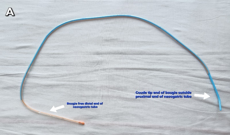
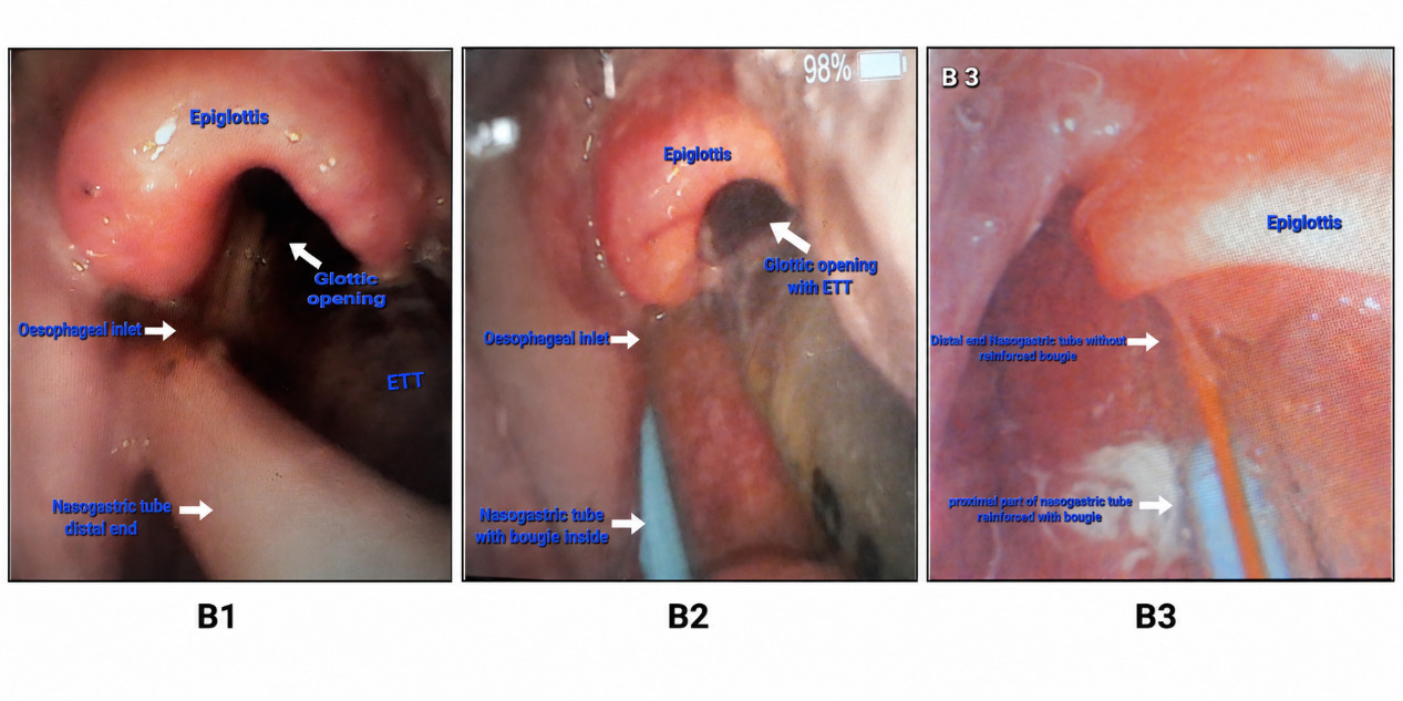
As a rescue measure, the first author modified a 16 FG (with outer diameter of 5.33mm and inner diameter of 3.3 mm), 105 cm Polymed PVC (Manufacturer: Polymedicure Limited) nasogastric tube by transecting it at 75 cm using a sterile blade and introducing straight end of adequately lignocaine gel lubricated PROACT® Pro-Breathe pediatric bougie (10 Fr, 3.3 mm external diameter, 60 cm, coudé tip) through its lumen till its hockey-stick angle; so that 10-15cm of NGT is soft at the distal end and not supported by the bougie (Figure 1A). Since the bougie fit snugly, the coudé tip angle also prevented distal migration of the bougie within the NGT. Under video laryngoscopic guidance, the nasally introduced NGT distal end was directed towards the oesophageal opening (Figure B1). At this point, the NGT, supported by the bougie, was gently advanced together (Figure B2). The bougie remained entirely within the NGT without protruding beyond its distal tip, while continuous control of its proximal end prevented distal migration. The added stiffness facilitated smooth oesophageal passage; we advanced the NGT to a depth of 65 cm, after which the bougie was withdrawn (Figure B3). Tube position in the stomach was initially assessed clinically by auscultation (because the patient was obese, the auscultation sounds were insufficient to confirm placement) and was subsequently confirmed by chest-upper-abdominal radiography. Prior lubrication of the bougie facilitated its easy withdrawal from the NGT while maintaining the inserted length. Around 10 cm of the nasogastric tube remained outside the nares and was secured to the nasal bridge using adhesive dressing tape. The proximal end of the tube was capped using its stopper. During feeding, the cap was removed, and the proximal end was connected to the hub of a feeding syringe as usual.
_with_the_bougie_inside.jpeg)
._b1__the_tip_of_the_ngt_is_in_the_esophagus._the_glottic_opening_is_visible_in_fron.png)
Keeping the bougie within the nasogastric tube and leaving 10–15cm of the distal tube unsupported was intended to reduce the risk of mucosal trauma, perforation, and false passage; however, these regular complications remain possible. No immediate or delayed complications were observed during the current procedure, including pharyngeal bleeding, oesophageal injury, aspiration, tube dysfunction, feeding intolerance, or the need for tube replacement during the remainder of the ICU stay.
Difficult NGT insertion is frequently encountered in perioperative and critical care practice. Several rescue techniques have been described, including ureteral guidewires, neck flexion with lateral neck pressure, external cricoid manipulation, and video laryngoscopy.1–3 Our technique is conceptually similar to guidewire-assisted methods but uses equipment routinely available in standard airway carts, permitting prompt bedside application. Endoscopic and fluoroscopic placement were not immediately available in our intensive care unit, making this approach a practical option for timely restoration of enteral nutrition.
This modified technique may represent a feasible rescue option after failure of conventional methods in mechanically ventilated patients. Contraindications include suspected pharyngeal or oesophageal perforation, oesophageal stricture, recent upper gastrointestinal surgery, caustic ingestion, and significant upper airway trauma. The procedure should be performed only under direct visualization by clinicians experienced in airway and upper gastrointestinal instrumentation, and should be abandoned immediately if resistance, bleeding, false passage, or loss of visualization occurs. As this is an off-label technique described in a single patient, it demonstrates only technical feasibility. Further studies are required before conclusions can be drawn regarding its safety, efficacy, or broader clinical applicability.
Funding
This study did not receive any specific grant from funding agencies in the public, commercial, or not-for-profit sectors.
Competing interests
All authors have completed the ICMJE uniform disclosure form and declare no conflict of interest.
Authorship
All authors meet the requirements of authorship and have reviewed and approved the final edit.
AI Statement
The authors confirm that no generative AI or AI-assisted technology was used to generate content.
Ethical Approval
Written informed consent for publication was obtained from the patient’s legally authorized representative prior to manuscript submission.